Flag findings that merit a closer look
Pancreatic lesions can be subtle on abdominal CT. PANCREASaver® provides an automated AI-assisted review that flags suspicious findings for radiologist attention.
PANCREASaver® is AI-powered, radiologist-reviewed medical device software for contrast-enhanced abdominal CT. It flags suspicious pancreatic findings for clinical review and does not issue an independent diagnosis.
U.S. status: FDA Pending; not yet cleared for U.S. marketing.
Meet PanCAD.ai at Booth 5132, McCormick Place, Chicago. Explore AI-assisted review of contrast-enhanced abdominal CT and discuss your clinical or integration needs.
Explore the event & schedule a demo ↗Built for the pressure points radiology teams face every day: subtle findings, crowded worklists and limited time.
Pancreatic lesions can be subtle on abdominal CT. PANCREASaver® provides an automated AI-assisted review that flags suspicious findings for radiologist attention.
Automatically analyzed studies can be flagged for review, helping teams focus attention where it matters most.
PANCREASaver® fits the PACS workflow your team already knows, adding decision support without requiring a new viewing habit.
From scan completion to AI analysis and radiologist review, this official overview shows how PANCREASaver® works alongside the imaging team.
For product and educational information; AI outputs are reviewed by healthcare professionals.
Watch on YouTube ↗
Pancreatic findings can be subtle on routine abdominal CT. PANCREASaver® analyzes the same images and flags areas for radiologist review within the existing workflow.
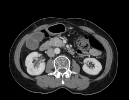
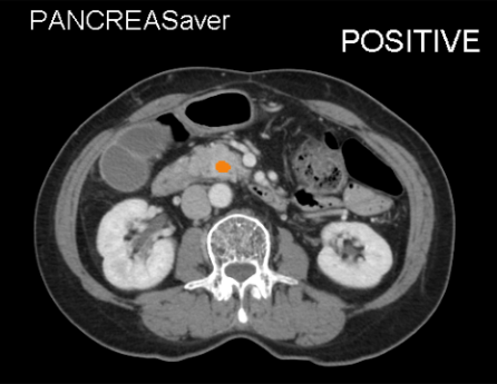
Real clinical CT images with actual PANCREASaver® output overlay. Radiologists always review and confirm AI findings.
PANCREASaver® is software as a medical device (SaMD) with FDA Breakthrough Device Designation. When an abdominal CT scan finishes, it analyzes the images, flags suspicious pancreatic findings, and returns results to the radiology workflow for review.
Analysis starts after the scan completes; the full pancreas is analyzed within minutes.
The cited study reported 92.1% sensitivity for lesions under 2 cm. PANCREASaver® is intended to support radiologist review, not replace clinical judgment.
Six peer-reviewed publications and a nationwide, multicenter AUC of 0.95, led by the National Taiwan University Hospital team.
The patient undergoes an abdominal CT; images are sent into the existing workflow.
PANCREASaver® analyzes the study within minutes; the radiologist reviews the findings.
Suspicious findings are marked and prioritized in the PACS worklist.
The radiologist reviews the findings and signs the final report; AI supports review and does not replace clinical judgment.
PANCREASaver® was designed around existing radiology workflows. It connects to your PACS/RIS environment; implementation details depend on the institution.
DICOM-native integration connects to the PACS/RIS infrastructure you already run — the software fits your existing environment.
Compatible with GE, Siemens, Philips, Canon and more — it runs on the scanners and viewers you already own.
Radiologists continue to review studies in their existing workflow while the software runs in the background and flags findings for attention.
Typically live in as little as 4 weeks with minimal IT lift — onboarding, training and go-live support included.
A DICOM link connects PANCREASaver® to your CT scanners and PACS.
The analysis starts automatically after the scan is complete; no separate application launch is required.
Suspicious studies are prioritized in the worklist with findings marked for radiologist review.
Structured findings are returned to your reporting workflow for review and sign-off.
PANCREASaver® integrates with clinical viewers and sign-in systems; it has been used in more than 5,500 patient cases across research and commercial applications.
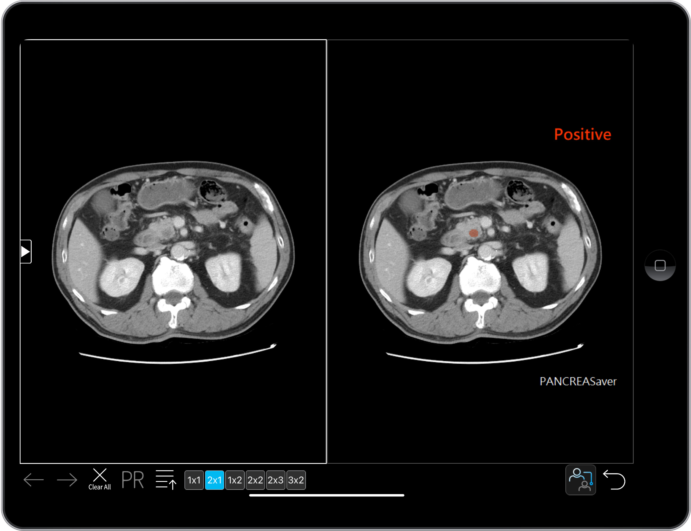 PANCREASaver®
Six papers in leading journals — every figure on this page carries a source you can open.
A cross-racial study of the AI detection engine reported 92.1% sensitivity for tumors under 2 cm and correctly recovered 11 of 12 cancers missed by radiologists. The study details and limitations are available in the original paper.
Nationwide multicenter performance with national-level AUC 0.95 — real clinical validation led by the NTUH team.
Cumulative use in more than 5,500 patient cases across research and commercial applications. Independent third-party validation is also part of the evidence base.
The full evidence hall — AUC curves, documented regulatory status, and the integration workflow.
Go to Clinicians →6 international journal papers with topics, abstracts and PubMed links.
Open the evidence →TFDA license, FDA Breakthrough Device Designation, FDA status, and 10 U.S. and Taiwan patents — stated exactly as they are.
See regulatory status →PANCREASaver® is AI-powered, radiologist-reviewed decision-support software for contrast-enhanced abdominal CT. It flags suspicious pancreatic findings for clinical review; it does not independently diagnose cancer or replace healthcare-professional judgment.
Yes. PANCREASaver® is DICOM-native and integrates with the PACS/RIS infrastructure you already run — no new viewer and no replacement of existing systems.
Typically live in as little as 4 weeks, with minimal IT lift. On-premises and cloud deployment options are available, and our team handles onboarding, training and go-live support.
No. Radiologists read exactly as they always have — AI runs automatically in the background and surfaces prioritized findings in the worklist.
It holds FDA Breakthrough Device Designation; FDA Pending. It is not yet cleared for U.S. marketing and is TFDA-licensed in Taiwan (license no. 衛部醫器製字第007946號).
Contrast-enhanced abdominal CT, per the authorized indications — the same scans your department already performs, with no extra acquisition protocol.
Verbatim third-party statements on the peer-reviewed studies behind PANCREASaver®. Quotes are about the research — not the product — and each carries its original source.
“
…accurate detection of pancreatic cancer on CT scans, especially for tumors smaller than 2 cm. Early detection… greatly increases the chances of survival.
“
This deep learning–based tool had very good accuracy, and sensitivity approaching that of subspecialist radiologists… Tools such as this promise to benefit routine workflow and patient care.
“
These promising results show that CNN as a second reader can reduce misdiagnosis of pancreatic cancer and possibly lead to improved patient outcomes.
Book a live demo to see how PANCREASaver® flags findings on clinical CT images.
Talk to us